

0 Products tested

Testing of Products
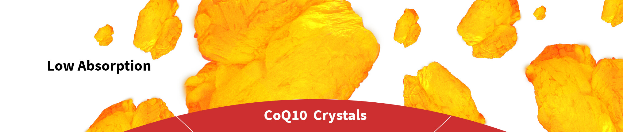
Different CoQ10 preparations have a very different appearance when viewed through a microscope. When we analyze a microscope image of a CoQ10 preparation we look for crystals - their size and shape. The CoQ10 raw material on which all CoQ10 preparations are based consists of large, irregular lumpy crystals. Unprocessed, this raw material will release only a few free CoQ10 molecules in the digestive tract.
In order to be absorbed from the gut, CoQ10 must be dissolved into free, single molecules. This is possible if the processing of the CoQ10 raw material produces small fragments with a high surface area-to-volume ratio like thin, needle-shaped crystals. Large lumpy crystals will only melt relatively few free CoQ10 molecules from their surface. Thin needle-shaped crystals are, however, able to melt completely at body temperature.
Hence, if a preparation with needle-shaped crystals is heated to body temperature (98.6°F) and looked at under a microscope, you will only see the oil and no crystal structures, as they will be completely dissolved.
A few preparations will show no crystals. It may mean two things: Either there is no CoQ10 in the preparation or that all CoQ10 is dissolved.
In this forum we only evaluate visible crystals at room temperature unless otherwise stated. The magnification range is 50X to 500X.
Types of crystals
 Polymorphic crystals
You could call them lumpy crystals. They will melt only a few free CoQ10 molecules from their surface after ingestion. It all comes down to the crystal's surface area-to-volume ratio.
 Acicular crystals
Are needle-shaped crystal. They are able to melt fully into single CoQ10 molecules at body temperature.
 Spherulite crystals
Are clusters of needle-shaped crystals radiating out from a center. They are also called snowflakes. They are able to melt fully into single CoQ10 molecules at body temperature.
 Plate-shaped crystals
Can be thick or thin. The thinner they are the better when it comes to dissolving at body temperature. Thick plate-shaped crystals will like lumpy crystals not melt completely at body temperature. Very thin plates might.
Microscopy Method
For this purpose of analysis Pharma Nord uses polarization microscopy. Polarization microscopes use a polarizing light to study objects, rather than natural or artificial light as with ordinary light microscopes. Polarization microscopy can provide some very detailed images. However, interference colors are formed, so the colors of images taken with a polarization microscope are not necessarily authentic as opposed to images taken with light microscopy.
When using polarization microscopy we try to get as three-dimensional images as possible. We do not use coverslides over the preparations, which is usually done using light microscopy. Furthermore, in some cases we use series of images with different depth focus which are subsequently united into a three-dimensional image.
It is also possible to evaluate crystals in CoQ10 preparations with other forms of microscopy.

| Label | Product data |
|---|---|
| Product name | Bio-Quinone Q10 Gold |
| Capsule | Softgel capsules |
| Oil | Soy oil |
| Batch | 1752121 |

Light microscopy of Q10 Gold taken by DTU. The image is collected at 50 times magnification

Polarization microscopy of Q10 Gold taken by Pharma Nord showing needle-shaped crystals and some fragmented spherulite crystals.

Polarization microscopy of Q10 Gold taken by Pharma Nord. Only needles and spherulites are seen.

Polarization microscopy of Q10 Gold taken by Pharma Nord. In the center a fragment of a spherulite crystal surrounded by needle-shaped crystals.

Polarization microscopy of Q10 Gold of recrystalization after heating showing only acicular crystals radiating out form a center core.

Polarization microscopy of Q10 Gold of recrystalization after heating showing a spherulite crystal dissolving. The first thing that happens is that the oil-condensing core melts from where needle-shaped Q10 crystals originates.

Polarization microscopy of Q10 Gold of recrystalization after heating showing spherulite and acicular crystals.

Polarization microscopy of Q10 Gold of recrystalization after heating showing only spherulite and acicular crystals. More needles are broken off and float freely before they begin to melt.
| Label | Product data |
|---|---|
| Product name | US001 |
| Capsule | Softgel capsules |
| Oil | Medium chain triglycerides |
| Expire | 07 - 2021 |

This picture focuses on a large spherulite crystal formation (a snowflake fragment) and also a mix of small polymorphic (lumpy) and plate-shaped crytals.

This picture shows a mix of plate-shaped and polymorphic (lumpy) crystals. At the top, center also a big dark lump of what look like densely packed needle-shaped crystals.

This picture is dominated by plate-shaped crystals. Top left is showing what might be part of a densely packed lump of needle-shaped crystals.
Here we have heated the oil. The needles have melted and reveals the content of polymorphic (lumpy) crystals.
| Label | Product data |
|---|---|
| Product name | US002 |
| Capsule | Soft gelatin capsules |
| Oil | Soy lecithin and medium chain triglycerides. |
| Expire | 09 - 2020 |

This is a low magnification overview.

At this magnification, scattered needles are seen

We see small polymorphic crystals, needles and a hint of plate-shaped crystals.

This picture is dominated by plate-shaped crystals and small polymorphic crystals.

A mix of spherulite fragments, needles and some small plate-shaped crystals.
| Label | Product data |
|---|---|
| Product name | US003 |
| Capsule | Softgel capsule |
| Oil | Soybean oil |
| Expire | 08-2021 |
 We see only polymorphic (lumpy) crystals in various sizes.

Magnification focusing on a single large lumpy crystal among many small ones.

This image shows a mixture of smaller lumpy crystals along with some plate-shaped crystals.